Download to read offline
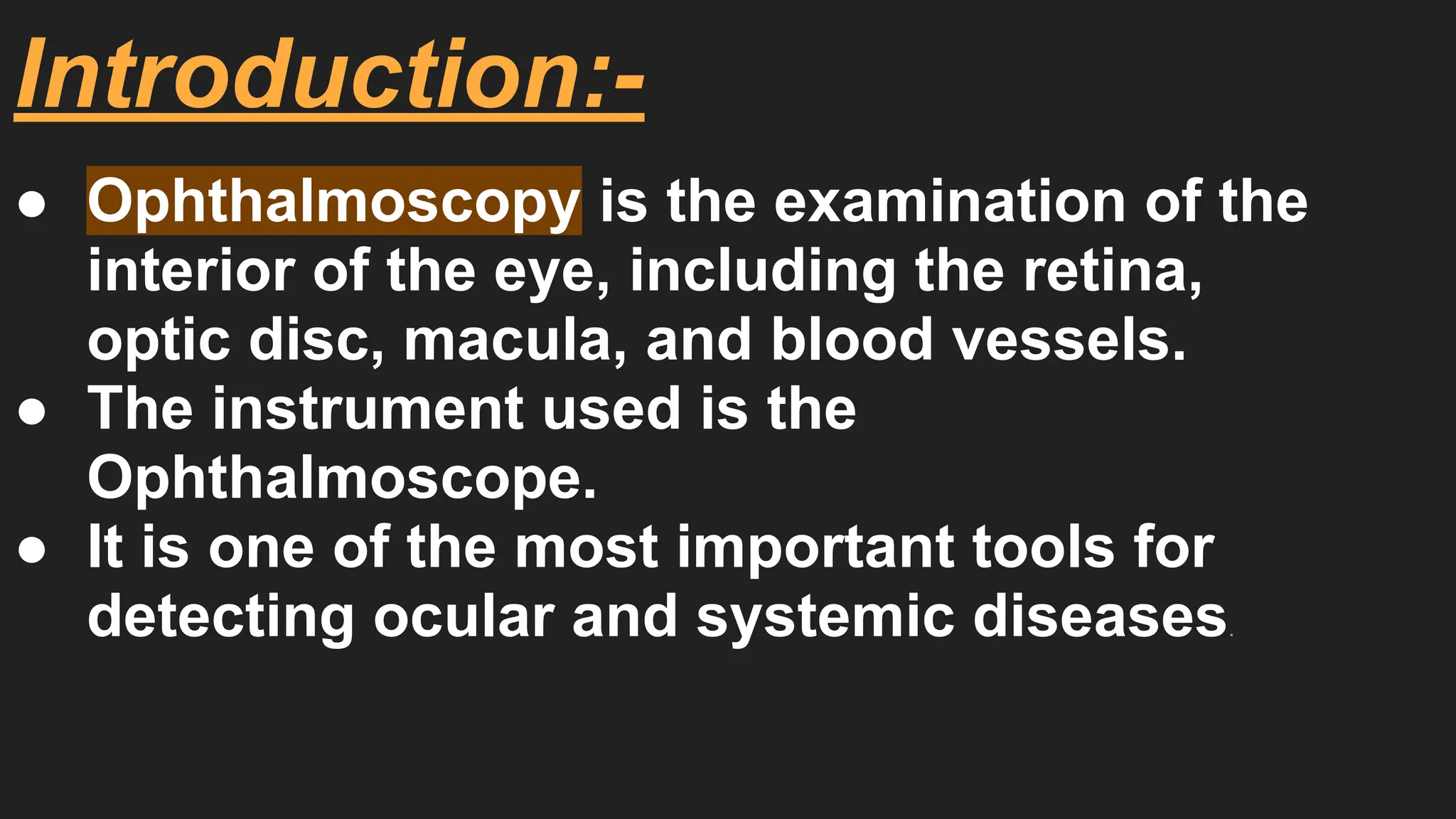

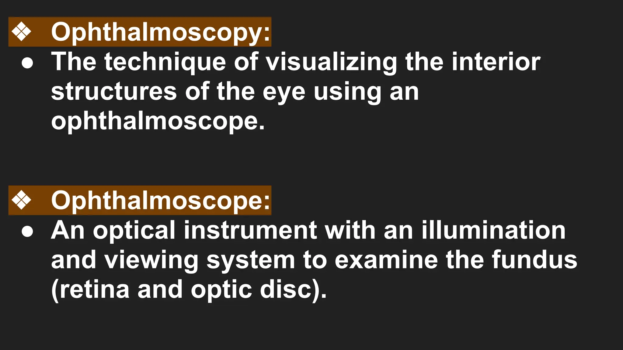
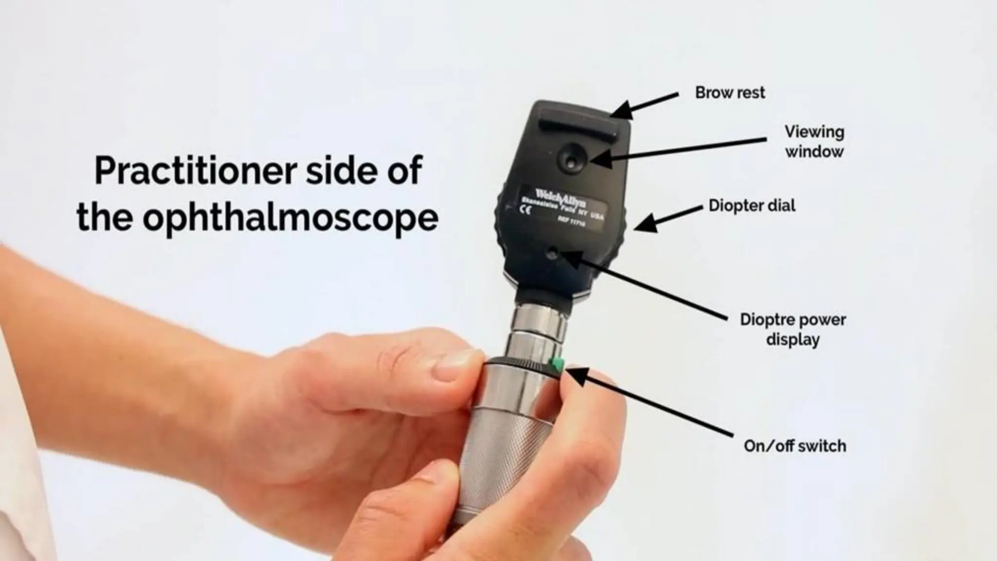

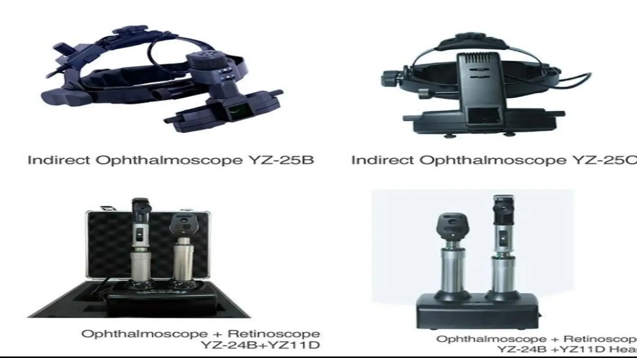
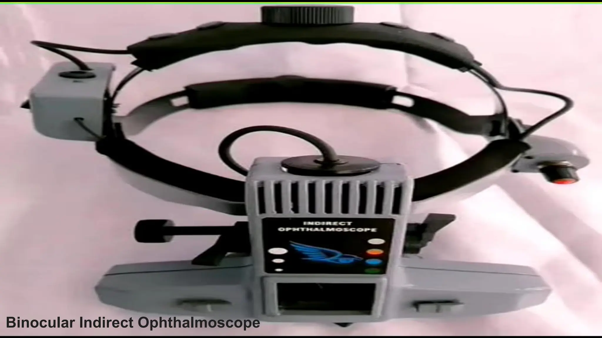
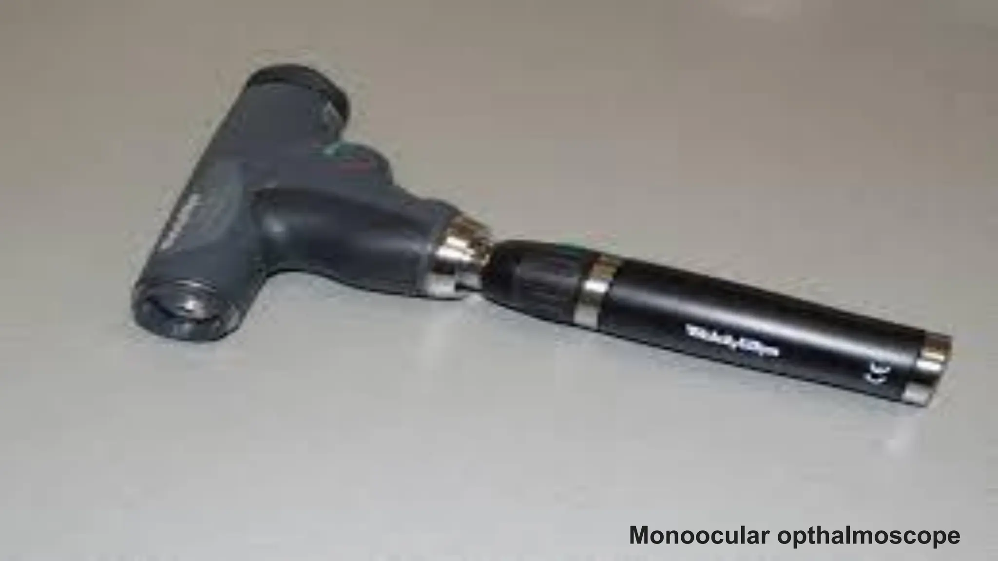
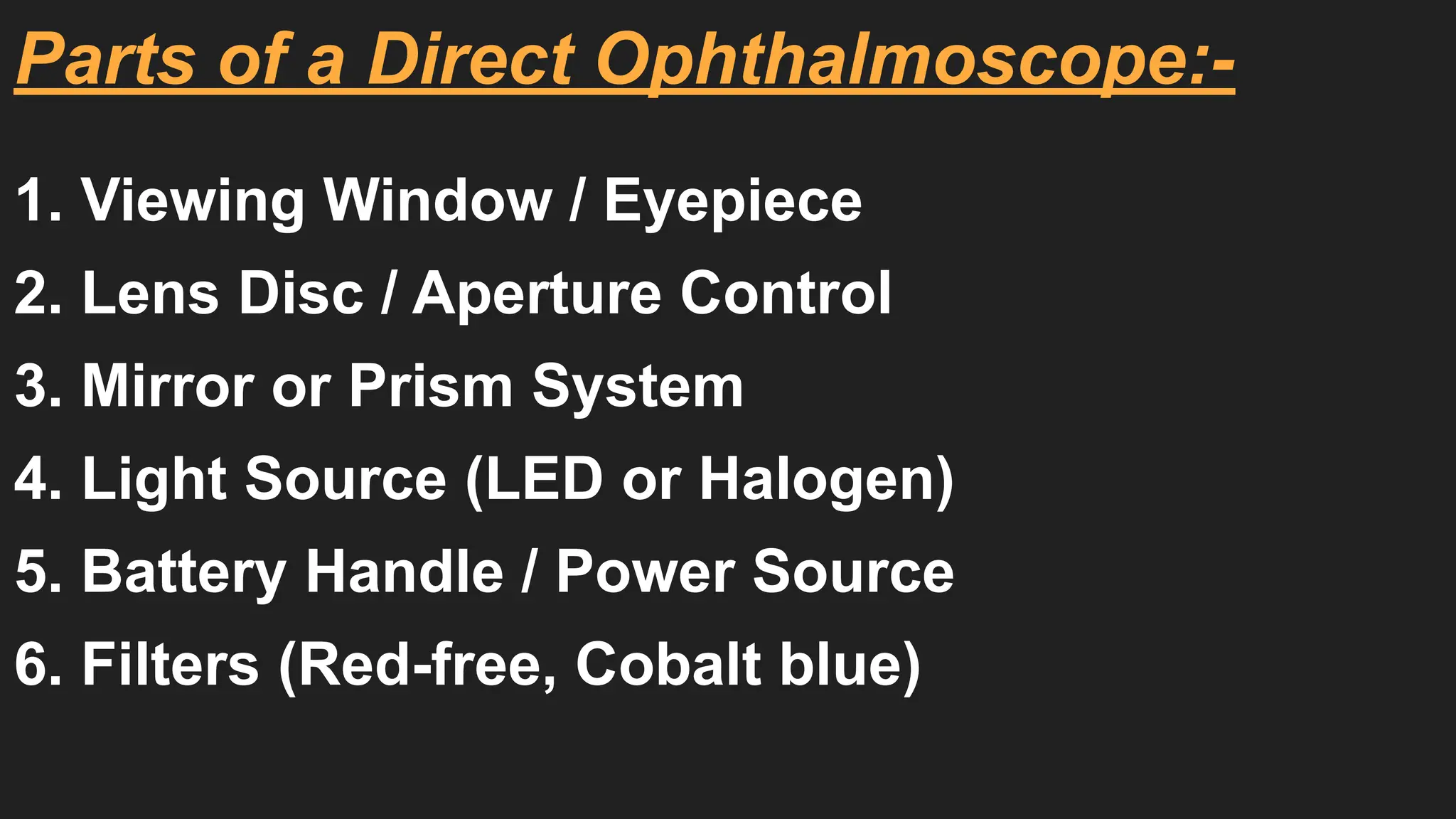
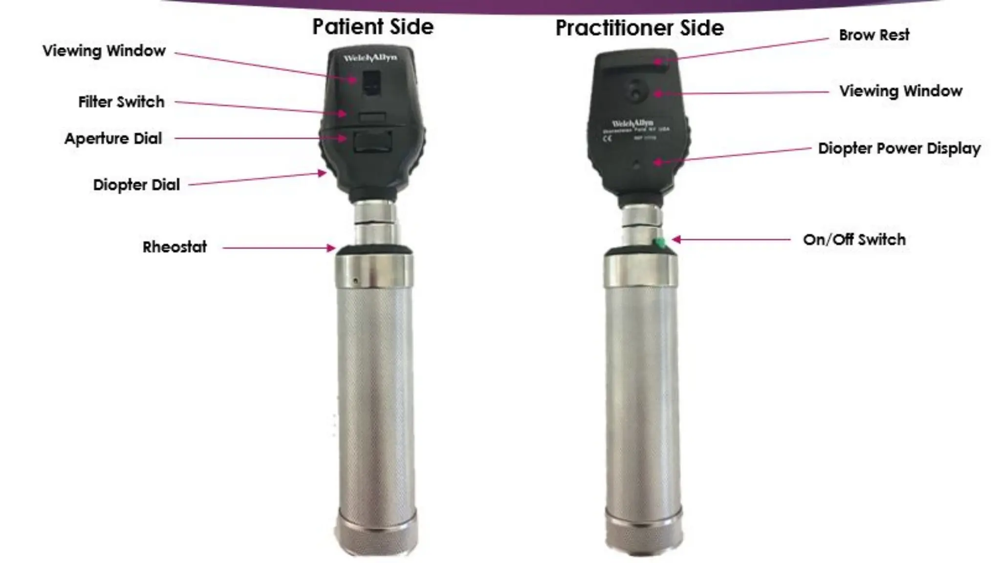
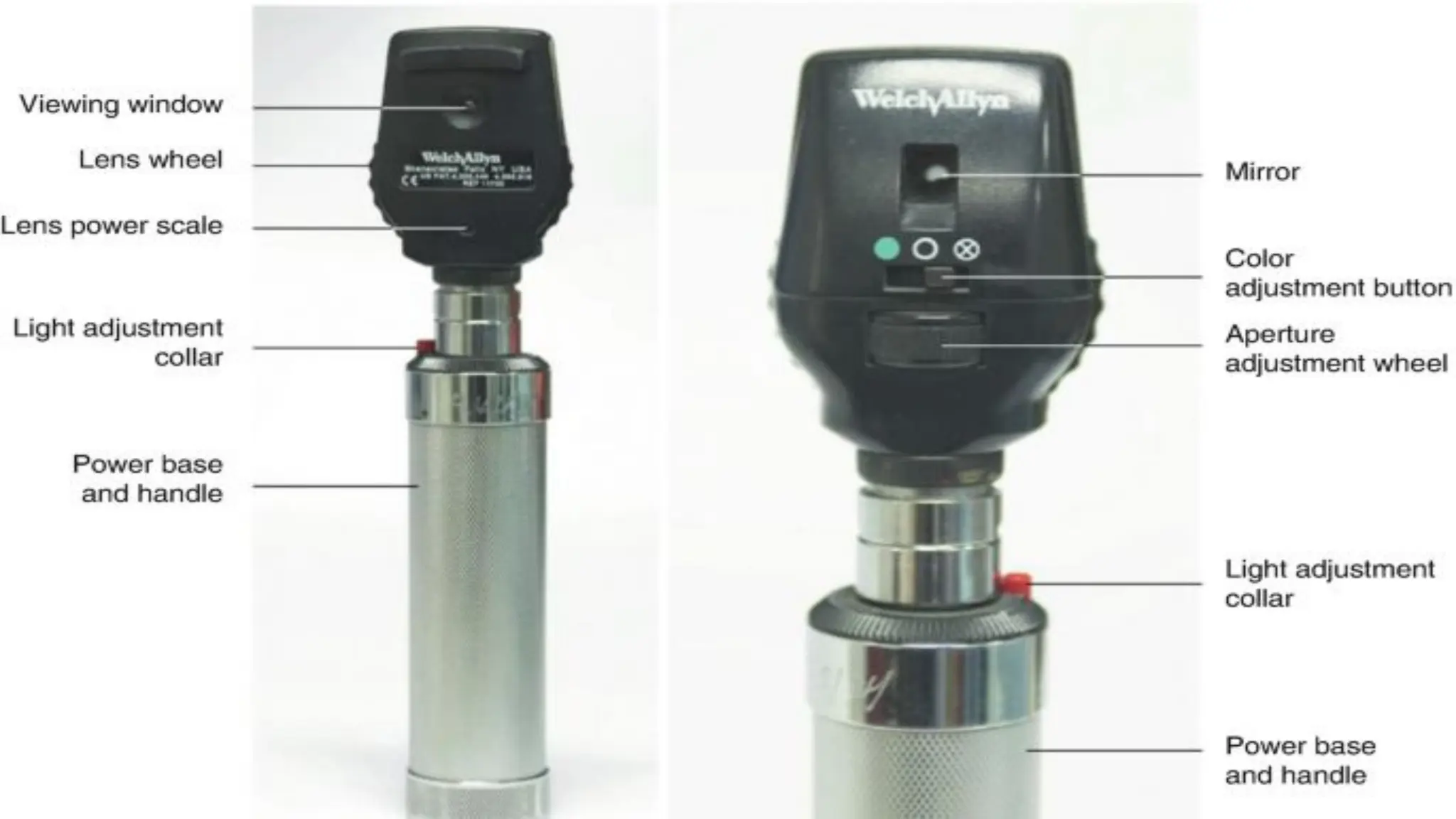
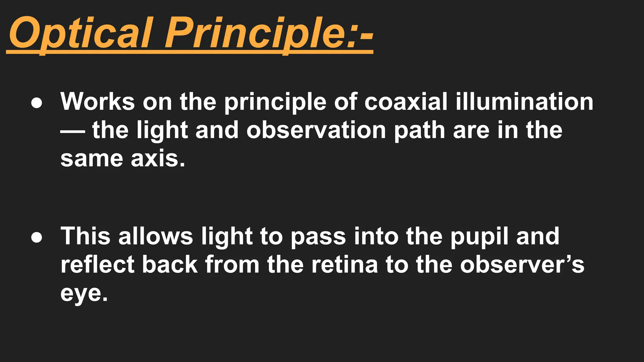
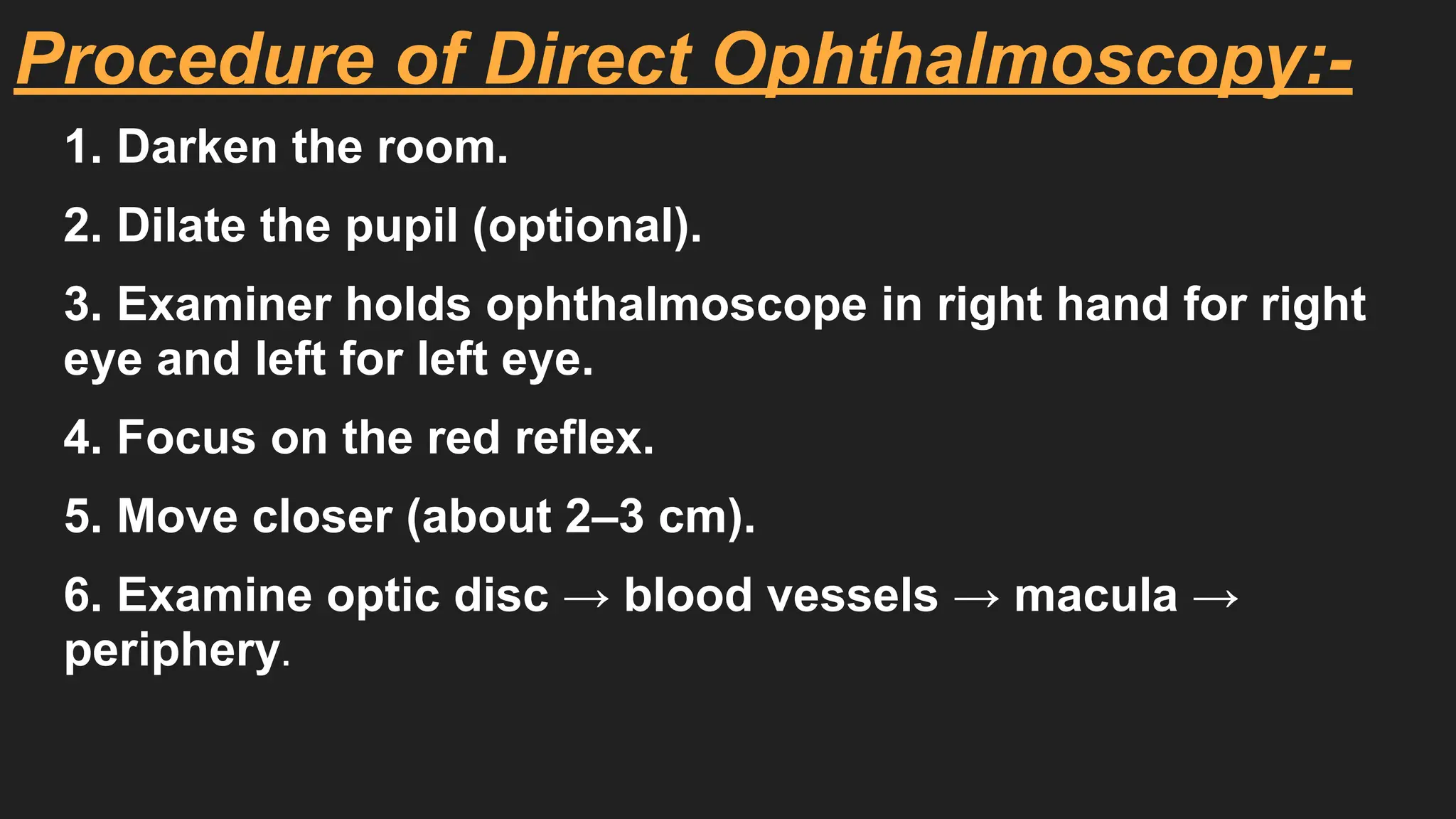
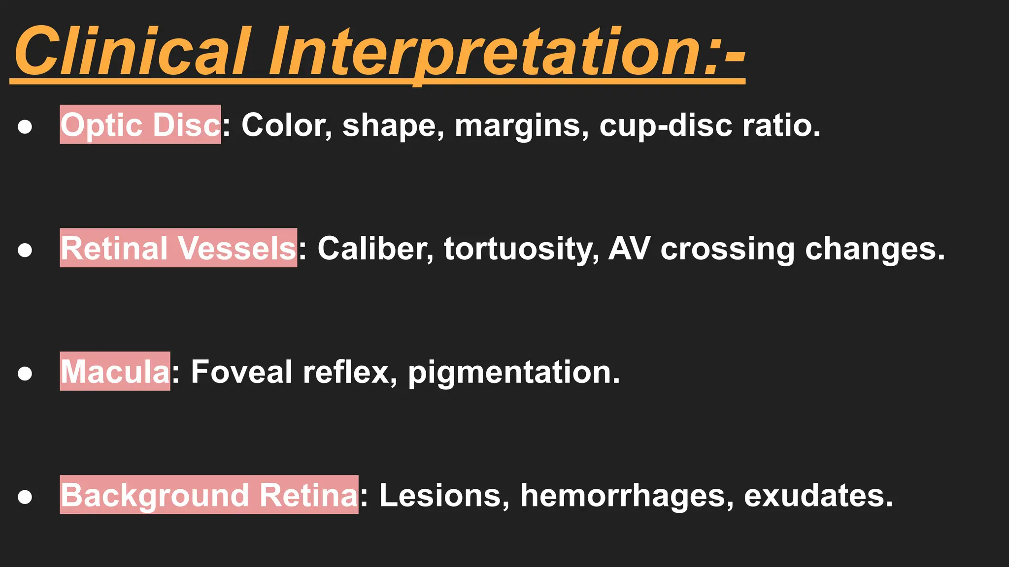
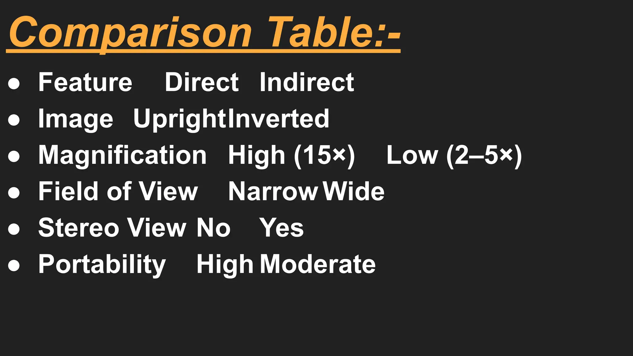
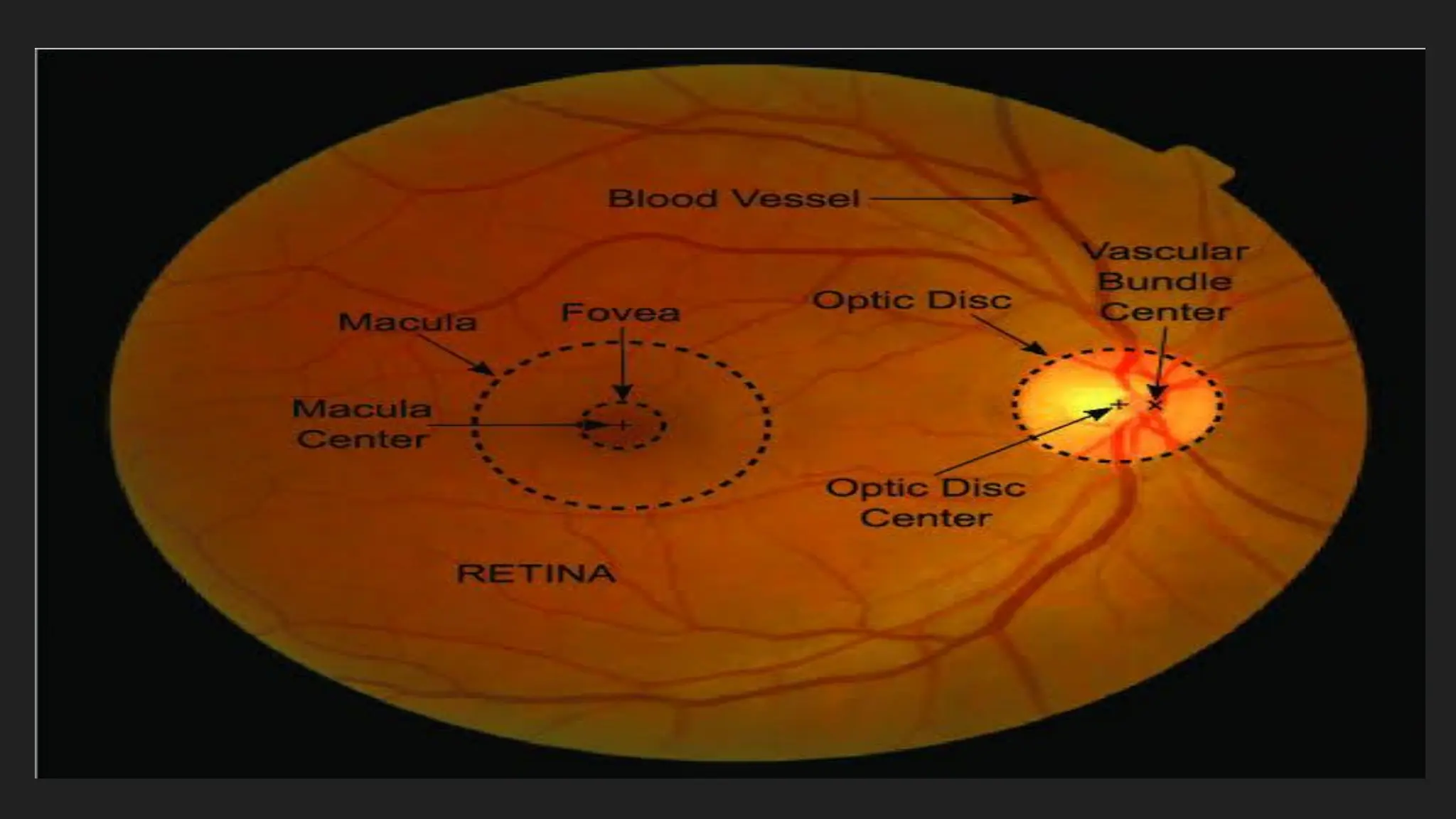
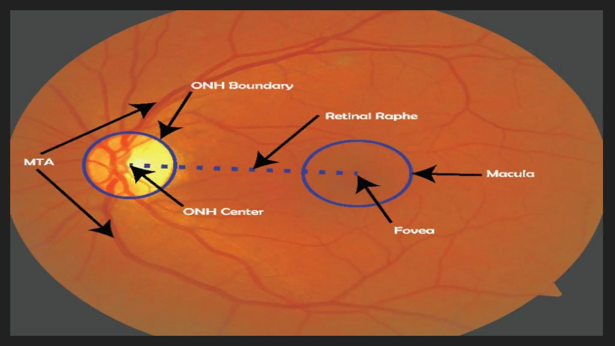
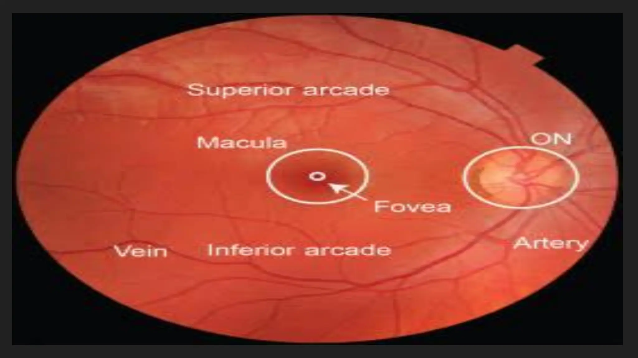
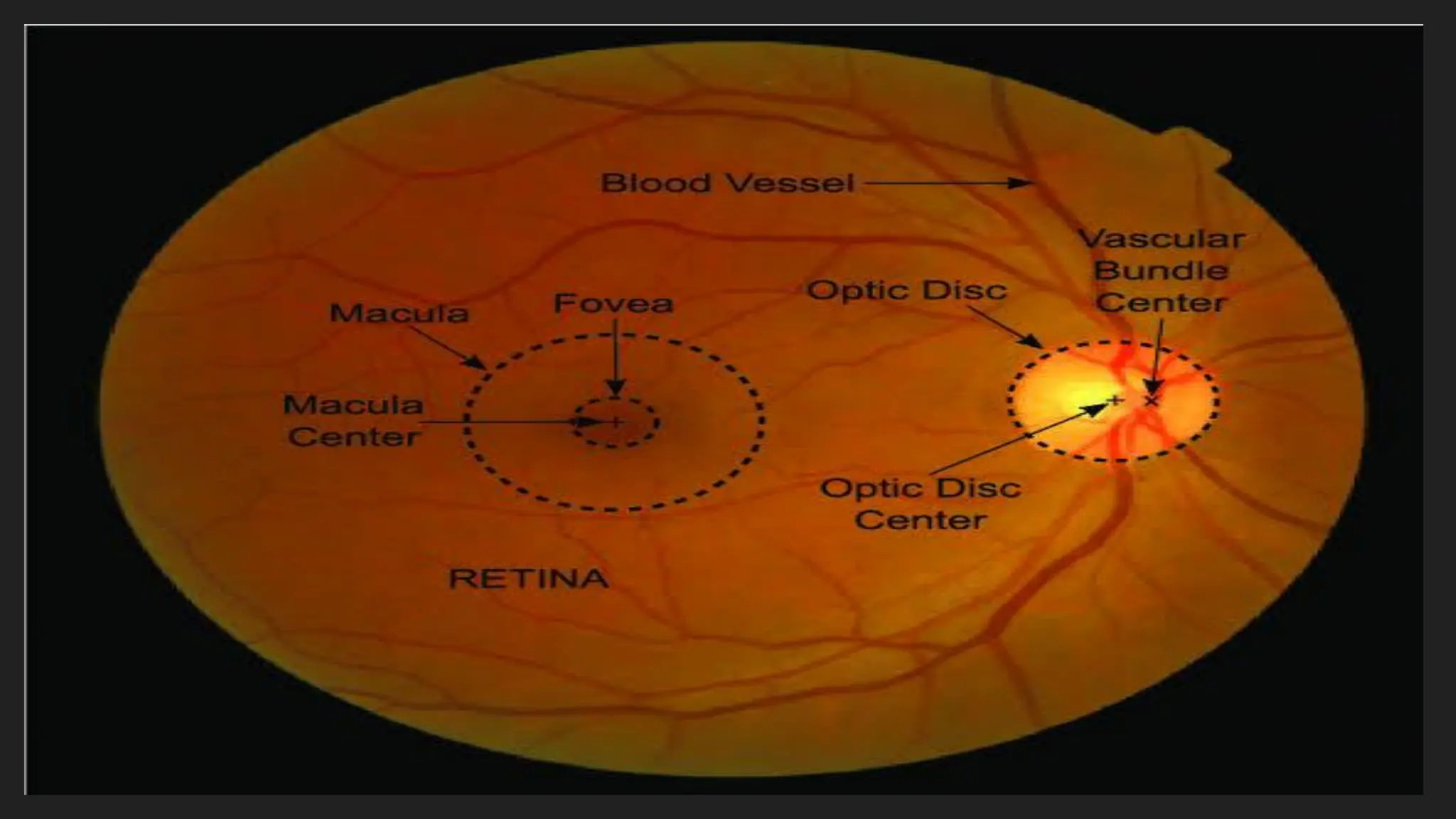


An ophthalmoscope is a medical instrument used by optometrists and ophthalmologists to examine the interior structures of the eye, especially the retina, optic disc, macula, and blood vessels. This procedure is known as ophthalmoscopy or fundoscopy. It is one of the most essential diagnostic tools in clinical eye examination because it helps in the early detection of many eye and systemic diseases such as glaucoma, diabetic retinopathy, hypertension, and papilledema. Principle: The principle of the ophthalmoscope is based on illumination and magnification. It uses a light source to illuminate the retina and a series of lenses to focus the image of the fundus for observation. The reflected light from the retina passes through the pupil and enters the examiner’s eye, allowing visualization of internal ocular structures. Types of Ophthalmoscope: 1. Direct Ophthalmoscope: It provides an upright and magnified (about 15x) image of a small area of the retina. It is compact, hand-held, and commonly used for routine eye checkups. The examiner views the fundus through a series of adjustable lenses and a light source aligned with the viewing aperture. Advantage: High magnification and portable. Disadvantage: Small field of view (about 5°). 2. Indirect Ophthalmoscope: It provides a real, inverted, and wider field of view (25–40°) of the retina. It uses a condensing lens (usually +20D) and a head-mounted light source. Advantage: Allows examination of the peripheral retina, useful for retinal detachment and vitreous hemorrhage. Disadvantage: Requires more skill and training. Parts of a Direct Ophthalmoscope: Head: Contains the bulb, viewing window, mirror, and lens disc. Handle: Contains the power source (battery or rechargeable unit). Lens Selector Wheel: Allows the examiner to adjust lenses from –40D to +40D for focusing. Aperture Selector: Includes different apertures such as small, large, slit, red-free (green), and fixation target for special observations. Uses of Ophthalmoscope: Examination of retina, optic disc, and blood vessels. Diagnosis of diabetic and hypertensive retinopathy. Detection of optic nerve diseases like optic atrophy and papilledema. Screening of glaucoma, macular degeneration, and retinal detachment. Used in neurological assessments to check for raised intracranial pressure. Advantages: Non-invasive and quick. Essential for early detection of systemic diseases. Portable and easy to use in clinical or community screening. Limitations: Small field of view in direct type. Requires clear ocular media (no corneal opacity or dense cataract). Conclusion: The ophthalmoscope is an indispensable diagnostic instrument in eye care, bridging the gap between clinical optometry and ophthalmology. Regular ophthalmoscopic examination helps in the early diagnosis and management of sight-threatening conditions and systemic diseases that manifest in the retina.Ophthalmoscope use, ophthalmoscope types, direct and


